
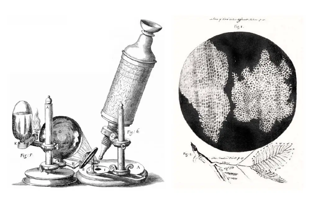
The cell is the basic unit or building block of living organisms. The cell was first observed and discovered under a microscope by Robert Hooke in 1665. The word “cell” came from Latin, which means “small room.” The cell membrane encloses the content of the cell and separates all biological activities from the outside world. Tiny structural parts inside the cell, called organelles, are involved in various specialized functions to keep the cell alive and active.
[In this figure] Left: The compound microscope used by Robert Hooke to discover “cells.” Right: Cell structure of cork illuminated by Robert Hooke in Micrographia, 1665.
Plants are multicellular organisms of the kingdom Plantae. Their features include:
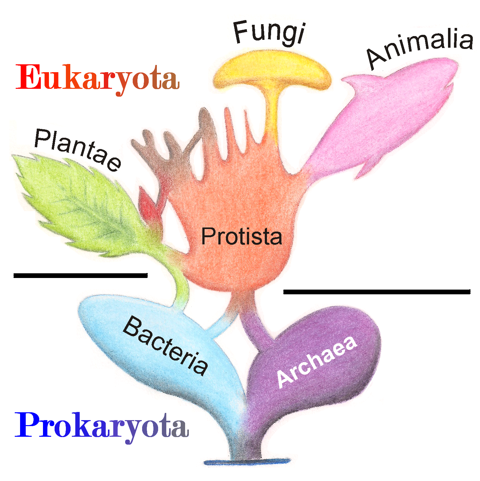
[In this figure] Tree of living organisms showing the origins of eukaryotes and prokaryotes.
Photo source: wiki.
Both animal and plant cells are classified as “Eukaryotic cells,” meaning they possess a “true nucleus.” Compared to “Prokaryotic cells,” such as bacteria or archaea, eukaryotic cells’ DNA is enclosed in a membrane-bound nucleus. These membranes are similar to the cell membrane, which is a flexible film of lipid bilayers. Eukaryotes also have several membrane-bound organelles. Organelles are internal structures responsible for various functions, such as energy production and protein synthesis.
Based on the current biological classification, both animals and plants are multicellular organisms, meaning that they consist of more than one cell. Different types of cells in a multicellular organism dedicate to different jobs.
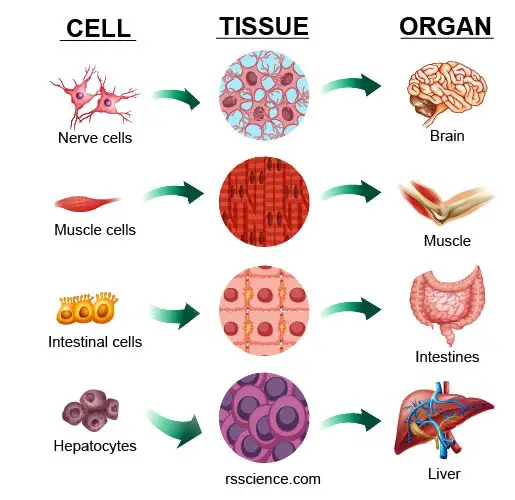
For example, cardiac muscle cells pump blood to circulate the body while intestinal cells absorb nutrients from the gut lumen into the bloodstream. Many cells assemble into a specific type of “tissue.” One or more tissues work together as an “organ.” Several organs join forces to carry out a specific physiological task and form a “system.”
There is a gray zone in the current biological classification, called Protista. The Protista, or Protoctista, is a kingdom of simple eukaryotic organisms, usually composed of a single cell or a colony of similar cells. A protist is not an animal, plant, or fungus. However, some protists may behave like animals or plants.
For example, protozoans are grouped as animal-like protists, and algae are referred to as mixed groups of plant-like protists. Interestingly, some species confuse the scientists by exhibiting both characteristics of animal and plant. The best example is Euglena, a single-celled microorganism that can harvest solar energy by its chloroplasts like a plant, but also swim around using its flagellum like an animal.
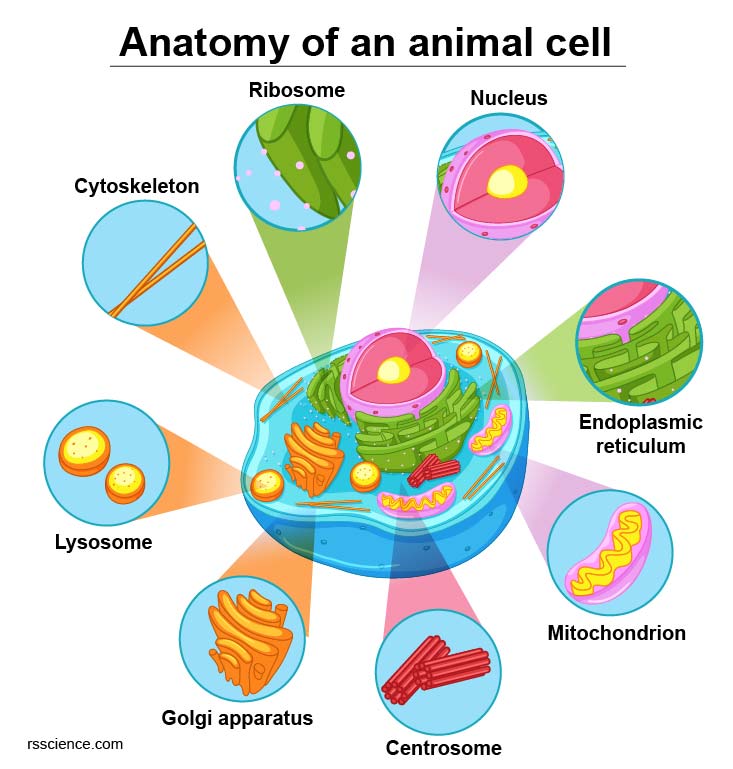
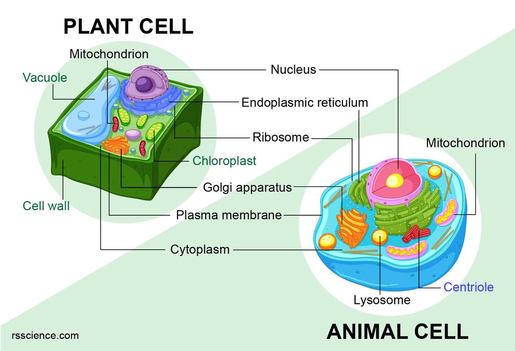
[In this figure] Diagram of an animal cell.

[In this figure] Diagram of a plant cell.
Like different organs within the body, animal and plant cells include various components known as cell organelles that perform different functions to sustain the cells as a whole. These organelles include:
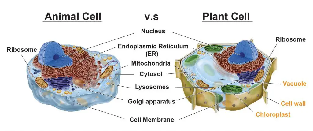
[In this figure] The cell anatomy of animal and plant cells.
The animal cell and plant cell share many organelles in common, such as a nucleus, ER, cytosol, lysosomes, Golgi apparatus, cell membrane, and ribosomes. The organelles unique for plant cells are vacuole, cell wall, and chloroplast (shown in orange text).
The most striking difference between animal cells and plant cells is that plant cells have three unique organelles: central vacuole, cell wall, and chloroplast. We summarize the major differences between plant and animal cells in this table.
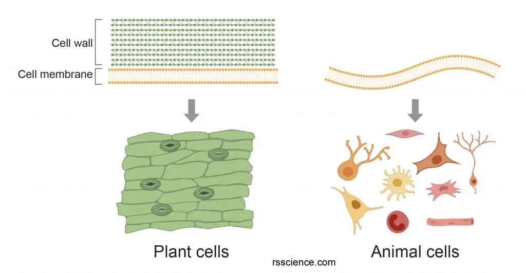
A difference between plant cells and animal cells is that plant cells have a rigid cell wall that surrounds the cell membrane. Animal cells do not have a cell wall. As a result, most animal cells are round and flexible, whereas most plant cells are rectangular and rigid. When looking under a microscope, the cell wall is an easy feature to distinguish plant cells.
[In this figure] Cell wall provides additional protective layers outside the cell membrane.
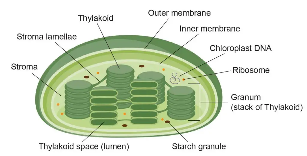
Plants are autotrophs, meaning they produce energy from sunlight through the process of photosynthesis. This function depends on the organelles called chloroplasts. Animal cells do not have chloroplasts. In animal cells, energy is produced from food (glucose) via a process of cellular respiration. Cellular respiration occurs in mitochondria in both animal and plant cells.
[In this figure] The structure of a chloroplast.
Plastids are double-membrane organelles that are found in the cells of plants and algae. Plastids are responsible for manufacturing and storing food. Plastids often contain pigments that are used in photosynthesis and different types of pigments can change the color of the cell. Chloroplasts are the most prominent type of plastids. Other plastids, like chromoplasts, gerontoplasts, and leucoplasts, may only occur in certain plant cells.
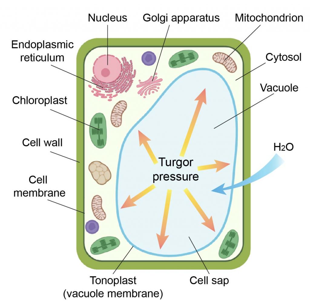
Animal cells have one or more small vacuoles, whereas plant cells have one large central vacuole that can take up to 90% of the cell volume. The function of vacuoles in plants is to store water and maintain the turgidity of the cell. Sometimes, vacuoles in plants also degrade cellular wastes like lysosomes. A layer of membrane, called tonoplast, surrounds the plant cell’s central vacuole. Due to the large size of the central vacuole, it pushes all contents of the cell’s cytoplasm and organelles against the cell wall. This may facilitate the cytoplasmic streaming of chloroplasts.
[In this figure] Drawing of a plant cell showing a large vacuole.
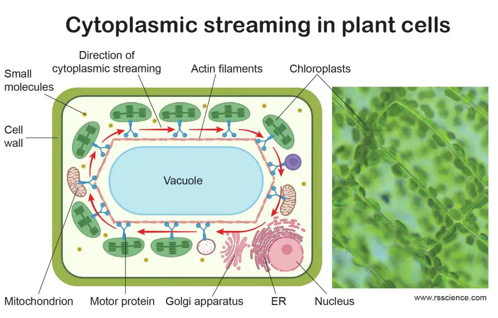
[In this figure] Cytoplasmic streaming in plant cells.
Cytoplasmic streaming circulates the chloroplasts around the central vacuoles in plant cells. This optimizes the exposure of light on every single chloroplast evenly, maximizing the efficiency of photosynthesis. The right image is the actual cytoplasmic streaming of chloroplasts in Elodea cells.
Created with BioRender.com
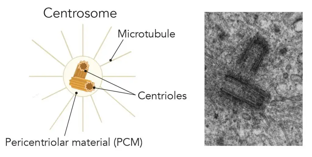
Centrioles are paired barrel-shaped organelles (centrosomes) located in the cytoplasm of animal cells near the nuclear envelope. All animal cells have centrioles, whereas only some lower plant forms have centrioles in their cells (e.g., the male gametes of charophytes, bryophytes, seedless vascular plants, cycads, and ginkgo).
[In this figure] Illustration and electron micrography of the centrosome.
Left: Centrosomes are composed of two centrioles arranged at right-angles to each other and surrounded by proteins called the pericentriolar material (PCM). Microtubule fibers grow from the PCM. Right: Electron microscopic images of centrioles. (Image: johan-nygren)
The lysosomes are small organelles that work as the recycling center in the cells. They are membrane-bounded spheres full of digesting enzymes. Lysosomes were considered to be exclusive to animal cells. However, this statement became controversial. Plant vacuoles are found to be much more diverse in structure and function than previously thought. Some vacuoles contain their own hydrolytic enzymes and perform the classic lysosomal activity like animals’.
Peroxisomes can be found in the cytoplasm of all eukaryotic cells, including both animal and plant cells. In plants, peroxisomes carry out two additional important roles.
First, peroxisomes (also called glyoxysomes) in seeds are responsible for converting stored fatty acids to carbohydrates, which is critical to providing energy and raw materials for the growth of the germinating plant. This occurs via a series of reactions termed the glyoxylate cycle.
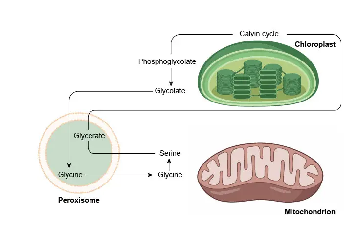
Second, peroxisomes in leaves are involved in the recycling of carbon from phosphoglycolate (a side product formed during photosynthesis) during photorespiration.
[In this figure] Photorespiration involves a complex network of enzyme reactions that exchange metabolites between chloroplasts, leaf peroxisomes, and mitochondria.
Plasmodesmata are microscopic channels that traverse the cell walls of plant cells and some algal cells, enabling transport and communication between them. Animal cells do not have plasmodesmata but have other ways to communicate between cells, like gap junctions or tunneling nanotubes (TNTs).
[In this figure] Plasmodesmata allow molecules to travel between plant cells through the symplastic pathway.
Photo source: wiki.
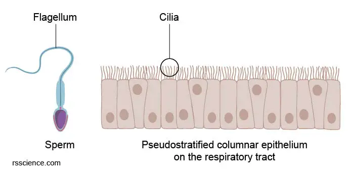
Two cellular structures that allow the movement of animal cells, flagella, and cilia (singular: flagellum and cilium), are absent in plant cells. Sperm cells are an excellent example of animal cells possessing flagella. Sperms use flagella for their movement toward the eggs. Cilia, on the other hand, act more like short hairs moving back and forth across the outside of the cell.
[In this figure] Cellular structures that allow the movement of animal cells: Flagellum (the tail of sperm) and Cilia (the waving hairs on the surface of airway cells).
You can easily find samples of animal and plant cells to look at under a microscope. See below to explore more:
Cheek cells (more specifically, epithelial cells) form a protective barrier lining your mouth. All you need to do is to gently scrape the inside of the mouth using a clean, sterile cotton swab and then smear the swab on a microscopic slide to get the cells onto the slide.
You can see our step-by-step guide, “Look at your cheek cells.“
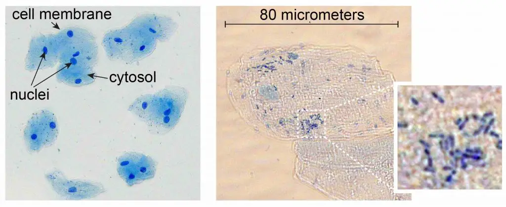
[In this figure] Cheek cells stained with Methylene Blue.
The left image is a low magnification. You can see the nuclei stained with a dark blue (because Methylene Blue stains DNA strongly). The cell membrane acts like a balloon and holds all the cell parts inside, such as a nucleus, cytosol, and organelles.
The right image is a high magnification. This check cell is about 80 micrometers in diameter. You can also see some small rod-shaped bacteria on the right image. Don’t worry; they are normal oral microbes.
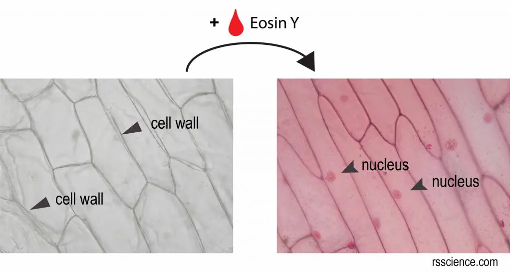
[In this figure] Microscopic view of onion skin.
The onion skin is a layer of protective epidermal cells against viruses and fungi that may harm the sensitive plant tissues. This layer of skin is transparent and easy to peel, making it an ideal subject to study plant cell structure. Without stains, you can only see the cell walls of onion cells. By staining Eosin Y, now you can see a nucleus inside an onion cell.
You can follow our step-by-step guide, “Look at the Plant Cells” to prepare your own onion skin slide.
In brief, the most striking difference between animal cells and plant cells is that plant cells have three unique organelles: central vacuole, cell wall, and chloroplast.
Animal cells have centrioles/centrosomes that most plant cells don’t. Some animal cells also have flagella and cilia, which are absent in plant cells.
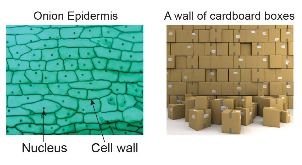
Due to the cell wall, many plant cells have a rectangular fixed shape.
[in this figure] The illustration of the cell wall.
The cell wall acts like a cardboard box that protects the soft cell membrane and cytoplasm. Like real cardboard boxes can be piled up to build a tall wall, the plant grows by adding cells one by one as living building blocks. The weight is loaded primarily on the structural cell walls.
Yes, plant cells have a layer of cell membrane underneath the cell wall. The cell membrane detaches from the cell wall under a hypertonic condition.
[In this figure] Turgor pressure on plant cells diagram.
Photo source: wiki.
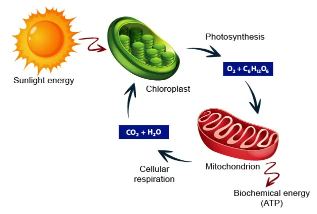
Yes, both animal and plant cells have mitochondria, but only plant cells have chloroplasts. In plant cells, chloroplasts absorb energy from sunlight and store it in the form of sugar (a process called photosynthesis). In contrast, mitochondria use chemical energy stored in sugars as fuels to generate ATP (called cellular respiration). Like animal cells, plant cells use ATP to drive other cellular activities.
[In this figure] The carbon cycle showing how energy flows between chloroplasts and mitochondria to benefit the ecosystem.
No, animal cells do not have a cell wall so they can freely change their cell shapes.
No, plant cells do not have centrioles for their mitosis except for some lower plant forms.
The presence of lysosomes in plant cells is under debate. Vacuoles in plant cells can fulfill the role of animal lysosomes.
Yes, plant cells have both free and endoplasmic reticulum-bound ribosomes for protein synthesis.
All cells (prokaryotic or eukaryotic; animal or plant) share four common components: (1) Plasma membrane, an outer covering that separates the cell’s interior from its surrounding environment.
(2) Cytoplasm, consisting of a jelly-like region within the cell in which other cellular components are found.
(3) DNA, the genetic material of the cell.
(4) Ribosomes, particles that synthesize proteins.
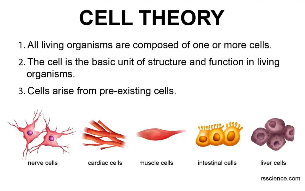
All cells on Earth have similar chemical compositions and meet the description of cell theory. The central dogma of molecular biology as “DNA makes RNA, and RNA makes protein” is also true in all cells.
Yes, both plants and animals are eukaryotes and have membrane-bound nuclei and organelles. Prokaryotic cells are bacteria and archaea.
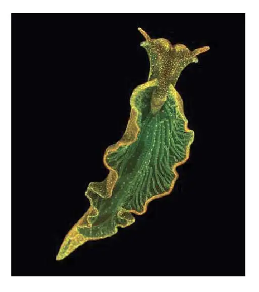
No, animals do not have chloroplasts, so they can not produce their food. However, some animals may borrow chloroplasts and live like a plant. Elysia chlorotica (common name the eastern emerald elysia) is one of the “solar-powered sea slugs,” utilizing solar energy to generate energy. The sea slug eats and steals chloroplasts from the alga Vaucheria litorea. The sea slugs then incorporate the chloroplasts into their own digestive cells, where the chloroplasts continue to photosynthesize for up to nine months.
[In this figure] Elysia cholorotica, a sea slug found off the U.S. East Coast, can steal photosynthetic chloroplasts from algae.
Photo source: Mary S. Tyler/PNAS
Yes, both plant and animal cells have a similar cytoskeleton. Constrained by the cell wall, the plant cell’s cytoskeleton does not allow a dramatic change of the cell shape. However, the cytoskeleton network of actin filaments, microtubules, and intermediate filaments generate shape, structure, and organization to the cytoplasm of the plant cell. The cytoskeleton also drives the cytoplasmic streaming in plant cells.
Cytokinesis occurs in mitosis and meiosis in both plant and animal to separate the parent cell from daughter cells.
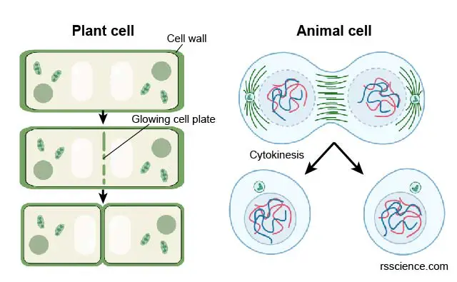
In plants, cytokinesis occurs when a cell wall forms in between the daughter cells. In animals, cytokinesis occurs when a cleavage furrow forms. This pinches the cell in half.
[In this figure] The difference of cytokinesis in plant and animal cells.